Papillary muscle abnormality as an electrocardiographic mimicker of apical hypertrophic cardiomyopathy: a case report
European Heart Journal - Case Reports


Abstract
Apical hypertrophic cardiomyopathy (ApHCM) is often associated with characteristic giant T wave inversions (GNT) in precordial leads without septal Q waves and increased QRS voltage on 12-lead electrocardiograms (ECGs). However, these electrocardiographic findings are not specific to ApHCM and can be mimicked by papillary muscle abnormalities. Differentiation between the two is important as the disease course, treatment, and prognosis differ substantially.
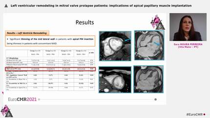
We report a case report of two such patients both of which presented with abnormal ECGs concerning for ApHCM. Echocardiogram did not show characteristic findings of ApHCM. Cardiac magnetic resonance imaging (MRI) showed apically displaced, hypertrophied papillary muscles responsible for electrocardiographic abnormalities.
Papillary muscle abnormalities including hypertrophy and/or apical displacement can result in giant negative T wave and increased QRS voltage like those seen in ApHCM and should be considered especially in otherwise healthy individuals with normal or near-normal transthoracic echocardiograms. Role of cardiac MRI is critical in this context and is the imaging modality of choice for accurate diagnosis.
Contributors

Jamal Janjua
Author
Joseph Burdowski
Author
Sumit Tickoo
Author
Ira Galin
Author
Edoardo Conte
Author
Diego Araiza-Garaygordobil
Author
Noldizsar Kovacs
Author
Gal Tsaban
Author
Nikesh Jathanna
Author
You may be interested in