C2. A Case of Arrhythmogenic Right Ventricular Cardiomyopathy Presenting with Recurrent Ventricular Tachycardia : The Role of Imaging to Confirm Diagnosis
European Heart Journal Supplements


Abstract
Arrhythmogenic right ventricular cardiomyopathy (ARVC) is an inherited condition which predominantly affects young people, characterized by fibrofatty replacement of the right ventricle. It is also commonly being underdiagnosed, so that the role of imaging is imperative to confirm diagnosis of ARVC patient.
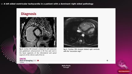
A 57-year-old man presented with palpitation, syncope and chest pain. The symptoms had begun since 3 month prior to hospital admission with no family history of premature sudden death. He had been hospitalized 3 times before due to palpitation. Electrocardiogram (ECG) revealed ventricular tachycardia with LBBB morphology and superior axis. Patient was undergoing 100 Joule cardioversion, and the ECG post-cardioversion showed sinus rhythm with inverted T-wave in inferior and anterior leads. Echocardiography revealed enlargement and reduced function of right ventricle (RV). Coroangiography revealed nonsignificant stenosis coronary artery disease. Cardiac MRI showed dyssynchrony RV contraction, reduced RV ejection fraction <40%, hyperintensity in RV endocardium and posteroinferior aspect of endocardium to myocardium left ventricle, representing the fatty infiltration. Our patient had 2 major criteria which established the diagnosis of definite ARVC according to revised Task Force Criteria. Patient was planned to have an implantable cardiac defibrillator.
Late clinical presentation of ARVC is rare, thus it should be included in the differential diagnosis when treating older patient with ventricular tachyarrhythmia. This case highlights the importance of imaging, either with echocardiography and cardiac MRI to confirm the diagnosis of ARVC as the potential cause sudden cardiac death.
Contributors

You may be interested in