Cardiac computed tomography and magnetic resonance imaging vs. transoesophageal echocardiography for diagnosing left atrial appendage thrombi
EP Europace Journal


Abstract
Transoesophageal echocardiography (TOE) is the gold standard for identification of left atrial appendage (LAA) thrombi. However, TOE is semi-invasive and cannot be performed in certain patients. Left atrial appendage thrombi can also be identified by cardiac computed tomography (CCT) and cardiac magnetic resonance imaging (CMR); however, the diagnostic performance of these techniques vs. TOE is unclear.
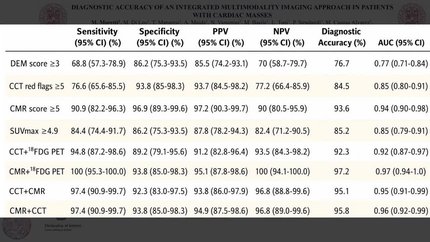
We performed a systematic review and meta-analysis of 22 CCT and 4 CMR studies comparing diagnostic performance to TOE for identification of LAA thrombi. Meta-regression was performed to determine whether expected sensitivity and specificity differed between early and delayed image acquisition protocols for CCT vs. TOE and between CCT and CMR. Cardiac computed tomography demonstrated sensitivity and specificity of 0.99 [confidence interval (CI 0.93–1.00)] and 0.94 (CI 0.90–0.97) respectively vs. TOE. A subgroup analysis comparing early vs. delayed protocol CCT imaging was performed showing no significant differences in sensitivity (
Cardiac computed tomography and CMR had good to excellent sensitivity and specificity vs. TOE. Further, there was no significant difference in the sensitivity and specificity of CCT vs. CMR, suggesting that both modalities can be considered reasonable alternatives to TOE in the identification of LAA thrombi. Cardiac magnetic resonance imaging may be especially beneficial when TOE and CCT cannot be performed.
Contributors

Tasnim Vira
Author
Petros Pechlivanoglou
Author
Kim Connelly
Author
Harindra C Wijeysundera
Author
Idan Roifman
Author
You may be interested in