In vivo assessment of aortic root geometry in normal controls using 3D analysis of computed tomography
European Heart Journal - Cardiovascular Imaging


Abstract
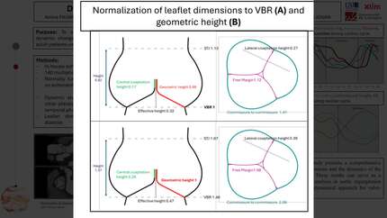
Understanding normal asymmetry in the aortic root could aid in the development of new surgical repair techniques or devices with improved haemodynamic performance. The purpose of this study was to assess geometric asymmetry and age-related changes in the normal aortic root using 3D computed tomography.
The institutional review board approved this retrospective study of 130 normal subjects (mean age, 51.4 years; 58 men). Specialized 3D software measured individual cusp sinus volumes (CSVs), cusp surface areas (CSAs), and intercommissural distances (ICDs). Age-related aortic root changes were evaluated with simple correlation, ANOVA test among age groups, and multivariable linear regression analyses. The CSV and CSA of left coronary cusp (LCC) were significantly smaller than those of right coronary cusp (RCC) and non-coronary cusp (NCC) (both,
The LCC was significantly smaller than the other two cusps, and the aortic root size increased with age.
Contributors

Dong Hyun Yang
Author
Dae-Hee Kim
Author
Mark D. Handschumacher
Author
Robert A. Levine
Author
Joon Bum Kim
Author
Byung Joo Sun
Author
Jeong Yoon Jang
Author
Namkug Kim
Author
Seunghee Baek
Author
Joon-Won Kang
Author
Jong-Min Song
Author
Duk-Hyun Kang
Author
Tae-Hwan Lim
Author

You may be interested in