Fusion imaging of pre- and post-procedural computed tomography angiography in transcatheter aortic valve implantation patients: evaluation of prosthesis position and its influence on new conduction disturbances
European Heart Journal - Cardiovascular Imaging


Abstract
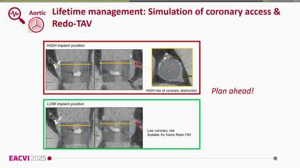
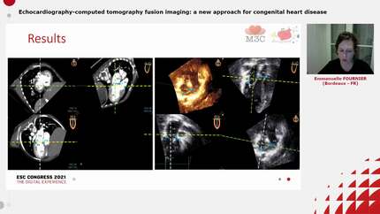
The purpose of this study was to evaluate prosthesis position by fusion of pre- and post-transcatheter aortic valve implantation (TAVI) computed tomography angiography (CTA) images and to investigate its influence on the occurrence of new conduction disturbances (CD).
We performed CTA fusion imaging in 120 TAVI patients (Edwards Sapien 3) on a standard image post-processing workstation to obtain a 3D reconstruction of the transcatheter heart valve (THV) position within the native annulus region. Optimal implantation depth of the THV was defined according to the manufacturers recommendations as 70–80% of the prosthesis above (aortic) and 20–30% below (ventricular) the native annulus plane. Pre- and post-interventional electrocardiograms (ECGs) were assessed for the development of new CD. THV position was found to be within, above, or below the prespecified margins in 32 patients (27%), 71 patients (59%), and 17 patients (14%), respectively. Interobserver reliability was high for fusion measurements [e.g. median THV position 0.983, 95% confidence interval (CI): 0.935–0.996]. Patients with low stent position were significantly more likely to develop new CD compared with patients with optimal or high stent position (
Fusion imaging of pre- and post-TAVI-CTA allows for the exact evaluation of THV position in relation to the native annulus plane. A low THV position as assessed by fusion imaging is associated with the development of new CD post-TAVI.
Contributors

Gregor Pache
Author
Jan Minners
Author
Manuel Hein
Author
Franz-Josef Neumann
Author
Philipp Breitbart
Author
You may be interested in