Impact of pectus excavatum on cardiac morphology and function according to the site of maximum compression: effect of physical exertion and respiratory cycle
European Heart Journal - Cardiovascular Imaging


Abstract
Previous studies have demonstrated diverse cardiac manifestations in patients with pectus excavatum (PEX), although mostly addressing morphological or physiological impact as separate findings. Using multimodality imaging, we evaluated the impact of PEX on cardiac morphology and function according to the site of maximum compression, and the effect of exertion and breathing.
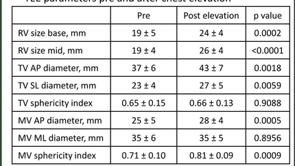
All patients underwent chest computed tomography, cardiac magnetic resonance (CMR), and stress echocardiography (echo) in order to establish surgical candidacy. We evaluated diastolic function and trans-tricuspid gradient during stress (echo); and systolic function and respiratory-related septal wall motion abnormalities (CMR). Patients were classified according to the site of cardiac compression as type 0 (without cardiac compression); type 1 (right ventricle); and type 2 [right ventricle and atrioventricular (AV) groove]. Fifty-nine patients underwent multimodality imaging, with a mean age of 19.5 ± 5.9 years. Compared with a sex and age matched control group, peak exercise capacity was lower in patients with PEX (8.4 ± 2.0 METs vs. 15.1 ± 4.6 METs,
The present study demonstrated that patients with PEX, particularly those with compression affecting the right ventricle and AV groove, manifest diverse cardiac abnormalities that are mostly related to exertion, inspiration, and diastolic function.
Contributors

Ignacio M Raggio
Author
Alejandro Deviggiano
Author
Gaston Bellia-Munzon
Author
Carlos Capunay
Author
Maximiliano Nazar
Author
Jorge Luis Martinez
Author
Patricia Carrascosa
Author
Marcelo Martinez-Ferro
Author
You may be interested in