Image fusion of integrating fluoroscopy into 3D computed tomography in guidance of left atrial appendage closure
European Heart Journal - Cardiovascular Imaging


Abstract
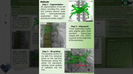
We evaluated the feasibility of left atrial appendage (LAA) closure guided by the image fusion of integrating fluoroscopy into 3D computed tomography (CT).
A total of 117 consecutive patients who underwent LAA closure with or without the image fusion were matched (1:2). Each LAA closure step of the Image fusion group was guided by the preprocedure CT and image fusion, especially in the plan of LAA measurement and transseptal puncture. All patients were successfully implanted with a WATCHMAN closure device. Comparing the two groups, the mean number of recapture times and the number of devices per patient of the Image fusion group were significantly lower (0.4 ± 0.5 vs. 0.7 ± 0.8,
Image fusion technique integrating fluoroscopy into the 3D CT is safe and feasible which can be easily incorporated into the procedural work-flow of percutaneous LAA closure. The fusion image can play an important alternative role in the plan of LAA measurement and transseptal puncture site for improving the LAA closure procedure.
Contributors

Bin-Feng Mo
Author
Yi Wan
Author
Abudushalamu Alimu
Author
Jian Sun
Author
Peng-Pai Zhang
Author
Ying Yu
Author
Mu Chen
Author
Wei Li
Author
Zhi-Quan Wang
Author
Yi-Gang Li
Author
You may be interested in