Fully automated machine learning-based selection of optimal bSSFP frequency offset for artifact reduction in cardiac MRI
European Heart Journal - Cardiovascular Imaging


Abstract
Type of funding sources: Private company. Main funding source(s): Research Support from Siemens Healthineers GmbH.
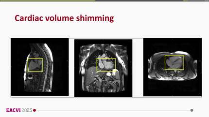
In bSSFP sequences commonly used for cardiac MRI, signal modulation (e.g. banding artifacts) due to B0 inhomogeneity is often observed, especially at higher field strengths. The spatial position of these artifacts can be shifted by a frequency offset to reduce artifacts in a region of interest (ROI), e.g. the heart. To this end, frequency scout (FS) scans are acquired to visually select the optimal frequency offset [1,2]. In this work, we propose a fully automated image-based system for selecting the optimal frequency offset on FS images based on machine learning.
The proposed prototype system consists of four main steps (
A total of 38 datasets, acquired on multiple clinical 3T MRI scanners (MAGNETOM Skyra, Vida, Prisma, Lumina; Siemens Healthcare, Erlangen, Germany), were used to evaluate the proposed system. All FS series were annotated manually and used to compare with the system output. The experts were allowed to select multiple possible optimal FS images within a FS series. In case of multiple annotations, the system output was labelled as correct when it selected one of the offsets chosen by the expert. Further, the generated weighting maps were visually evaluated.
The proposed system achieved an accuracy of 92.1% compared to experts’ ground truth annotations. From the failed cases (n=3), the maximum difference was off by 2 frames. Based on the generated weighting maps, a reasonable decision on the selection of the optimal frequency offset is made. The algorithm successfully selects an FS image with minimized banding and flow artifacts within the ROI (
Initial results demonstrate the feasibility of the proposed system to automatically select the optimal frequency offset on FS scans. Therefore, it can improve the automation of a cardiac MRI workflow. An example of the result of each step
Contributors

You may be interested in