Impact of right ventricular reserve on exercise capacity and quality of life in patients with left ventricular assist device
European Heart Journal


Abstract
Right heart failure following left ventricular assist device (LVAD) implantation is a major complication which significantly impairs functional capacity and quality of life (QoL). Right ventricular (RV) reserve function may limit exercise capacity and QoL in LVAD patients; however, most patients show normal RV haemodynamics at rest.
The aim of this study was to investigate whether RV reserve assessed by the changes of RV function during exercise is correlated with exercise capacity and QoL in patients with LVAD.
We prospectively examined 20 consecutive LVAD patients who were admitted to our university hospital between June 2020 and November 2021 after excluding those who were unable to perform exercise (n=8). All patients underwent invasive exercise right heart catheterisation with simultaneous echocardiography in the supine position. RV stroke work index (RVSWI) was calculated as 0.0136 × stroke volume index × (mean pulmonary artery pressure [mPAP] − right atrial pressure [RAP]) at rest and during exercise. Exercise capacity and QoL were assessed by 6-minute walk distance (6MWD) and peak oxygen consumption (VO2) in cardiopulmonary exercise testing, and the EuroQol visual analogue scale (EQ-VAS), respectively. The patients were divided into two groups according to the median ΔRVSWI (RVSWI change from rest to peak exercise) of 1.45 (interquartile range [IQR] −0.31 to 8.25) g/m2.
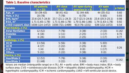
Patients were predominantly male (75%) and the median age was 47 (IQR 38–60) years. Patients with lower ΔRVSWI had significantly higher change on RAP (P=0.019), but significantly lower change on mPAP (P<0.001) compared to those with higher ΔRVSWI. There were no significant differences in age, gender, primary aetiology of heart failure, type of LVAD devices, or echocardiographic parameters including tricuspid annular plane systolic excursion, and RVSWI at rest between the groups. ΔRVSWI during exercise were positively correlated with 6MWD (R=0.69, P<0.01) and peak VO2 (R=0.66, P<0.01) (Figure A). In addition, ΔRVSWI during exercise were positively correlated with the EQ-VAS (R=0.48, P=0.031). On the other hand, there was no significant correlation between RVSWI at rest and 6MWD (R=−0.11, P=0.63) and peak VO2 (R=0.13, P=0.95), and the EQ-VAS (R=0.11, P=0.61). During a median follow-up period of 312 (IQR 176–369) days, adverse events occurred in 3 patients (15%), including 1 death and 2 hospitalisations for major bleeding and right heart failure. Kaplan-Meier analysis revealed that the adverse events more frequently occurred in patients with lower ΔRVSWI compared to those with higher ΔRVSWI (Figure B).
ΔRVSWI was positively correlated with 6MWD, peak VO2 and EQ-VAS irrespective of RV function at rest. Our findings suggest that the assessment of RV reserve function using ΔRVSWI would be useful for risk stratification in patients with LVAD.
Type of funding sources: None.
Contributors

S Takenaka
Author
T Sato
Author
T Nagai
Author
K Omote
Author
K Kamiya
Author
T Konishi
Author
Y Kobayashi
Author
A Tada
Author
Y Mizuguchi
Author
Y Takahashi
Author
S Naito
Author
K Saiin
Author
S Ishizaka
Author
S Wakasa
Author
T Anzai
Author
You may be interested in