The impact of ventricular-arterial coupling on aerobic capacity across the heart failure spectrum
European Heart Journal


Abstract
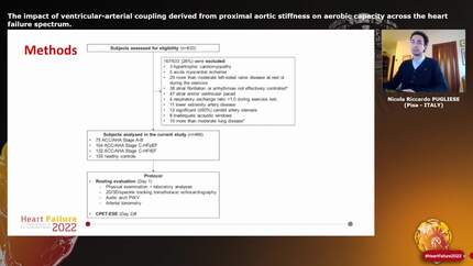
Ventricular-arterial coupling (VAC) can be evaluated as the ratio between arterial stiffness (pulse wave velocity, PWV) and myocardial deformation (global longitudinal strain, GLS).
To evaluate VAC across the spectrum of heart failure (HF).
We introduced a Doppler-derived, single-beat technique to estimate aortic arch PWV (aa-PWV) in addition to tonometry-derived carotid-femoral PWV (cf-PWV). We measured PWVs and 3D-GLS in 155 healthy controls, 75 subjects at risk of developing HF (American College of Cardiology/American Heart Association Stage A-B) and 236 patients in HF Stage C with preserved (HFpEF, n=104) or reduced ejection fraction (HFrEF, n=132). We evaluated peak oxygen consumption (VO2) and peripheral extraction (AVO2diff) using combined cardiopulmonary-echocardiography exercise stress.
aa-PWV was obtainable in all subjects and significantly lower than cf-PWV in all subgroups (p<0.01). PWVs were directly related and increased with age (all p<0.0001). cf-PWV/3D-GLS was similarly compromised in HFrEF (1.08±0.36) and HFpEF (1.05±0.22), while aa-PWV/3D-GLS was more impaired in HFpEF (0.69±0.11) than HFrEF (0.60±0.15; p<0.01). Stage A-B had values of cf-PWV/3D-GLS and aa-PWV/3D-GLS (0.66±0.25 and 0.47±0.12) higher than controls (0.47±0.10 and 0.40±0.10) but lower than Stage C (all p<0.01). Peak AVO2diff was inversely related with cf-PWV/3D-GLS and aa-PWV/3D-GLS (all p<0.01). cf-PWV/3D-GLS and aa-PWV/3D-GLS independently predicted peak VO2 in the overall population (adjusted R2=0.32 and 0.35; all p<0.0001) but only aa-PWV/3D-GLS was independently associated with flow reserve during exercise (R2=0.51; p<0.0001).
Abnormal VAC is directly correlated with greater severity of HF and worse functional capacity. HFpEF shows a worse VAC than HFrEF when expressed by aa-PWV/3D-GLS.
Type of funding sources: None.
Picture 1
Contributors

N R Pugliese
Author
A Balletti
Author
S Armenia
Author
N De Biase
Author
F Faita
Author
A Mengozzi
Author
F Paneni
Author
F Ruschitzka
Author
A Virdis
Author
L Ghiadoni
Author
S Taddei
Author
B Williams
Author
F Antonini-Canterin
Author
S Masi
Author
You may be interested in