Is cardiac magnetic resonance of additional help in premature ventricular contraction work-up?
European Heart Journal


Abstract
Premature ventricular contractions (PVCs) are common in routine medical practice. PVCs are commonly asymptomatic but can also result in palpitations, dyspnea, presyncope or PVC-induced cardiomyopathy that may lead to sudden death. Cardiovascular magnetic resonance (CMR) provides morphological, functional, and tissue characterization in a single setting. CMR imaging proved to reliably identify fibrotic lesions and, hence, has developed into a valuable tool for etiological orientation in patients with PVCs.
The aims of this study were: 1) to evaluate which ventricular arrythmia (VA) characteristics predicted CMR abnormalities and 2) the diagnostic yield and the impact in the management of CMR-based imaging versus non-CMR-based imaging in patients referred for PVCs.
This monocentric retrospective study was conducted in patients with PVCs referred for CMR imaging. Their baseline interview and characteristics, physical examination, surface electrocardiogram (ECG) analysis, ambulatory monitoring, echocardiography and treadmill exercise test data were collected when they were available. The CMR (1,5 T and 3.0 T) protocol included cine imaging and late gadolinium enhancement (LGE) using standard cardiac geometries.
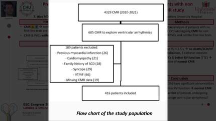
We analyzed 168 patients (65% males, mean age 54.2±16.8 years) referred to our center for PVCs over a two-year period. CMR reported abnormal findings in 73 patients (43.5%) with the diagnosis of structural heart disease (SHD) in 46 patients (27.4%). Left-ventricular late gadolinium-enhancement was documented in 43 patients with abnormal CMR. Using a multivariate analysis, age (p=0.026), sustained ventricular tachycardia (p=0.002) and presence of cardiomyopathy on echocardiography (p<0.001) significantly predicted the presence of abnormal findings on CMR. Compared to non-CMR-based imaging, CMR had a diagnostic yield, by modifying the presumed diagnosis or by finding abnormalities requiring follow-up in 73 patients (43.5%).
Age, sustained ventricular tachycardia and presence of cardiomyopathy on echocardiography were the strongest predictors of positive CMR imaging results. Although echocardiography is generally used in first intention for the diagnosis of cardiomyopathy in patients with PVCs, this study showed that CMR-based imaging has a robust diagnostic yield in this indication.
Type of funding sources: None.
Contributors

You may be interested in