Hepatic T1-time predicts cardiovascular risk in all-comers referred for cardiovascular magnetic resonance
European Heart Journal


Abstract
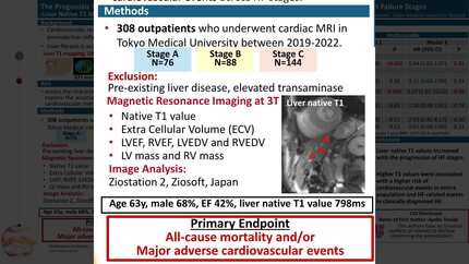
Liver damage is frequently observed in patients with cardiovascular disease (CVD) but infrequently quantified. We hypothesized that in patients with CVD undergoing cardiac magnetic resonance (CMR), liver T1-times indicate liver damage and are associated with cardiovascular outcome.
We measured hepatic T1-times, displayed on standard cardiac T1-maps, in an all-comer CMR-cohort. At the time of CMR, we assessed validated general liver fibrosis scores. Kaplan-Meier estimates and Cox-regression models were used to investigate the association between hepatic T1-times and a composite endpoint of non-fatal myocardial infarction, heart failure hospitalization, and death.
1022 participants (58±18 y/o, 47% female) were included (972 patients, 50 controls). Hepatic T1-times were 590±89ms in patients and 574±45ms in controls (p=0.052). They were significantly correlated with cardiac size and function, presence of atrial fibrillation, NT-pro-BNP levels, and gamma-glutamyl-transferase levels (p<0.001 for all). During follow-up (58±31 months), a total of 280 (29%) events occurred. On Cox-regression, high hepatic T1-times yielded a significantly higher risk for events (adj.HR 1.66 [95% CI: 1.45–1.89] per 100ms increase, p<0.001), even when adjusted for age, sex, left and right ventricular ejection fraction, NT-proBNP, and myocardial T1-time. On restricted cubic splines, we found that a hepatic T1-time exceeding 610ms was associated with excessive risk.
Hepatic T1-times on standard CMR scans were significantly associated with cardiac size and function, comorbidities, natriuretic peptides, and independently predicted cardiovascular mortality and morbidity. A hepatic T1-time >610ms seems to indicate excessive risk.
Type of funding sources: None.
Figure 1
Contributors

K Mascherbauer
Author
C Dona
Author
M Koschutnik
Author
V Dannenberg
Author
C Nitsche
Author
F Duca
Author
D Beitzke
Author
C Loewe
Author
E Waldmann
Author
M Trauner
Author
P Bartko
Author
G Goliasch
Author
J Mascherbauer
Author
C Hengstenberg
Author
A Kammerlander
Author
You may be interested in