Poster No. 142 An unusal heart-kidney connection: from diagnosis to staging of renal cell carcinoma
Cardiovascular Research


Abstract
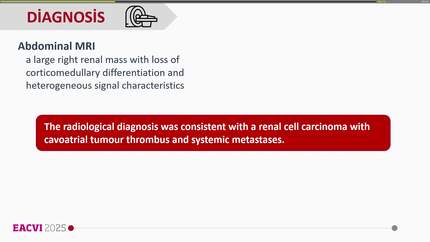
A 58-years-old man is admited in emergency department with complaints of 2-days le flank pain and hematuria. When questioned, he mentions 6-months bilateral low back pain. His medical history was relevant for tobacco consumption (80 pack-year) and acute myocardial infarction in 2011. During abdominal examination, a painless 7 cm mass is detected in the left abdominal flank. Blood tests were remarkable for macrocytic anemia, increased D-dimers and serum calcium levels and leukocytosis. A thoracoabdominal-pelvic CT was performed to exclude advanced neoplasia. A 15×13 cm heterogeneous renal lesion involving the lower half of the left kidney was shown. This lesion extended through the renal vein and suprarenal inferior vena cava (IVC) to the right atrium. Bilateral pulmonary thromboembolism and metastatic lesions in the liver, left adrenal gland, latero-aortic ganglia and lung parenchyma were also detected. A thrombus was confirmed in the IVC with extension to the Eustachian valve by echocardiography. Metastatic renal carcinoma was assumed and enoxaparin was started. A renal biopsy identified clear cell renal carcinoma (CCRC). Embolization of the left renal artery was performed and Palliative Care follow-up was started after multidisciplinary team discussion. Renal cell carcinoma accounts for 2% of all tumors in adulthood with its classic symptomatic triad (flank pain, hematuria, and palpable abdominal mass) occuring only 9% of cases, as does endovascular dissemination from the renal vein and IVC. It is associated with a median survival of 5 months. We emphasize the importance of complete clinical examination and the recognition of poor prognosis predictors of CRCL.
Contributors

Jéni Quintal
Author
Paula Lopes
Author
Joana Póvoa
Author
Aissato Cassama
Author
Ana Cristina Diogo
Author
Vanessa Pereira
Author
You may be interested in