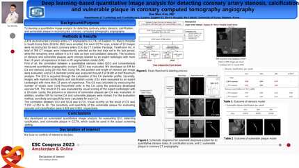
Poster No. 071 Application of artificial intelligence in coronary CT angiography:a potential gatekeeper strategy?
Cardiovascular Research


Abstract
Medical artificial intelligence (AI) is rapidly developing and moving from the research field to daily clinical practice. AI algorithms have demonstrated high performance and computational efficiency, reducing the degree of manual input and processing time.
This study aimed to determine the impact of an AI-enabled coronary computed tomography angiography (CCTA) analysis for comprehensive evaluation in patients (P) with suspected coronary artery disease (CAD).
We analysed 100 CCTA exams from a cohort of symptomatic P with mild-to-moderately abnormal non-invasive ischemia test. Stenosis severity was assessed by level III experts (manual evaluation, MEv). A novel AI-based software tool (automatic evaluation, AEv) was also used to quantify coronary stenosis and characterize plaque phenotype. In P later referred for invasive coronary angiography (ICA), diagnostic and revascularization yields of MEv and AEv were compared.
100P, 52% male, mean age 68 ± 10 years, one-third had typical angina. Prevalence of obstructive CAD determined by MEv and AEv was 25% and 21%, respectively, with a significant association between both assessments (
Based upon MEv, referring physician decided to proceed to ICA in 22P (21P with significant stenosis). For those undergoing ICA,13P also had obstructive CAD established by AEv. Diagnostic yields for MEv and AEv-guided ICA was 82% and 60%, and revascularization yields 73% and 60%, respectively.
AEv atherosclerosis quantification revealed significant differences between P who did not undergo ICA, P referred for ICA without significant stenosis and P with obstructive CAD on ICA: total (126 vs. 312 vs. 518 mm3,
A diagnostic strategy using AI-based analysis of coronary stenosis severity on CCTA had a similar performance compared to MEv. In addition, risk prediction can be enhanced by AI assessment of plaque composition. This study is an example of the potential role of AI in the CCTA workflow.
Contributors

José Miguel Viegas
Author
João Ferreira Reis
Author
Rita Teixeira
Author
Tiago Mendonça
Author
Ruben Ramos
Author
Sofia Jacinto
Author
Hugo Marques
Author
Luísa Figueiredo
Author
James Earls
Author
Rui Cruz Ferreira
Author
You may be interested in