Progressive morphological changes of a bioprosthetic mitral valve on serial chest CT preceding severe haemodynamic deterioration in systemic sclerosis: a case report
European Heart Journal - Case Reports


Abstract
Structural valve deterioration is an important cause of bioprosthetic valve dysfunction. Morphological abnormalities may precede clinically significant haemodynamic deterioration, but recognition of this progression may depend on longitudinal comparison of imaging studies.
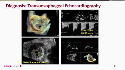
A 76-year-old man with a surgical bioprosthetic mitral valve implanted in 2014 was diagnosed with systemic sclerosis-associated interstitial lung disease in 2022 and subsequently underwent serial chest computed tomography (CT). Echocardiography in 2022 and March 2023 demonstrated preserved prosthetic valve haemodynamics. In August 2024, he was admitted with progressive heart failure and severe bioprosthetic mitral stenosis. Retrospective review of serial CT demonstrated progressive leaflet changes, from a relatively thin appearance in 2022 to mild thickening in 2023 and marked diffuse thickening with an apparent fusion-like configuration in May 2024. Transoesophageal echocardiography demonstrated severe leaflet thickening and restriction with a suspected superimposed thrombotic component. Despite therapeutic enoxaparin, recurrent pulmonary oedema prompted redo mitral valve replacement. Operative examination demonstrated diffuse leaflet thickening, rigidity, and commissural fusion, with non-dominant pannus and thin thrombotic material.
This case illustrates the value of longitudinal comparison of existing imaging studies. Progressive morphological changes of a bioprosthetic valve were retrospectively identified on chest CT performed for a non-cardiac indication before severe haemodynamic deterioration was recognized by echocardiography. This does not support routine CT surveillance; rather, abnormalities encountered on existing extracardiac imaging may provide an additional signal to consider earlier cardiac reassessment.
Contributors

Phu D Bui
Author
Anh T Duong
Author
Huy Q Dang
Author
Christoph Sinning
Author
Deepti Ranganathan
Author
You may be interested in