Highly vascularized left atrial myxoma mimicking a cardiac haemangioma: a case report
European Heart Journal - Case Reports


Abstract
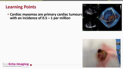
Primary cardiac tumours are rare; atrial myxomas predominate, while haemangiomas represent only 1%–2.8%. Differentiating them is challenging when hypervascular features overlap.
An 81-year-old woman presented with a large, hypervascular left atrial mass supplied by the circumflex artery. Following uneventful surgical resection, histopathology confirmed a cardiac myxoma despite imaging suggesting a haemangioma.
Hypervascularity cannot reliably distinguish myxomas from haemangiomas. Although multimodal imaging is important for surgical planning, definitive diagnosis relies strictly on histopathological evaluation.
Contributors

Juan Manuel Aragón González
Author
Henry Noel Fajardo Perdomo
Author
Felipe Rodríguez Mora
Author
Antonio González Calle
Author
Takeshi Kitai
Author
Issameddine Ajmi
Author
Liang Zhang
Author
Deepti Ranganathan
Author
You may be interested in