Cardiac hydatidosis: enigmatic presentation of enigmatic diagnosis: a case report
European Heart Journal - Case Reports


Abstract
In endemic countries, hydatid disease is often overlooked because of its non-specific clinical presentation and the limited use of multimodality imaging. Unusual anatomical locations and atypical presentations further complicate diagnosis. Multimodality imaging not only helps to confirm the diagnosis but also plays a crucial role in treatment planning.
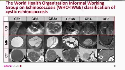
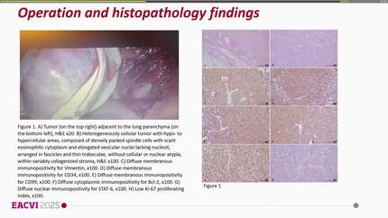
A 45-year-old man from a rural area presented with a 2-year history of haemoptysis and exertional dyspnoea. He had received antitubercular therapy for 1 year without improvement. Subsequent computed tomography angiography at a secondary care centre suggested pulmonary thromboembolism, and he was referred to our centre for further evaluation. He also had a history of foul-smelling cough and peripheral eosinophilia, without any identifiable risk factors for pulmonary thromboembolism. Routine echocardiography revealed a large multi-cystic mass attached to the basal interventricular septum and protruding into the right ventricular outflow tract. Hydatid disease was strongly suspected and subsequently confirmed by serological testing and transoesophageal echocardiography. Cardiac computed tomography and magnetic resonance imaging were performed to better define the nature, location, and extent of the lesion. The patient was treated with albendazole followed by uneventful surgical excision and remains asymptomatic on follow-up.
This case illustrates a rare presentation of cardiac hydatidosis masquerading as pulmonary embolism. Multimodality imaging was essential in characterizing the lesion, defining its anatomic extent, and guiding management. Surgical excision can be performed safely when carefully planned, and perioperative chemotherapy reduces the risk of recurrence.
Contributors

Aditya Kapoor
Author
Shantanu Pande
Author

Konstantinos Stathogiannis
Author
Jan Henzel
Author
Rami Riziq Yousef Abumuaileq
Author
Alessandro Palmieri
Author
Deepti Ranganathan
Author
You may be interested in