The normal D-dimer paradox: pulmonary artery intimal sarcoma masquerading as pulmonary embolism: a case report
European Heart Journal - Case Reports


Abstract
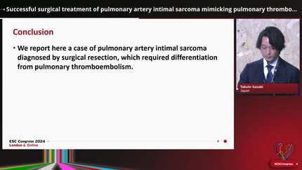
Pulmonary artery intimal sarcoma is an exceptionally rare and aggressive malignancy that can closely mimic pulmonary embolism. Overlapping clinical and imaging features frequently lead to diagnostic anchoring and delayed recognition, with devastating prognostic consequences.
A 50-year-old woman presented with a nine-month history of progressive dyspnoea and cough and was admitted with hypotension, jugular venous distension, peripheral oedema, and systolic murmurs. Laboratory testing revealed a D-dimer level of 0.5 μg/mL (reference range 0.1–0.6 μg/mL). Transthoracic echocardiography showed right ventricular dilatation and a mass involving the pulmonary valve and main pulmonary artery. Computed tomography pulmonary angiography demonstrated an expansile lesion of the main pulmonary artery extending into both branches, with extraluminal involvement and new pulmonary nodules. Despite therapeutic anticoagulation for presumed pulmonary embolism, there was no clinical or radiological improvement. Histological diagnosis was obtained through biopsy of a breast lesion, revealing a high-grade sarcoma with vascular differentiation consistent with pulmonary artery intimal sarcoma. Given extensive local invasion and poor functional status (ECOG 4), curative treatment was not feasible, and the patient died 30 days after definitive diagnosis.
In the present case, pulmonary artery intimal sarcoma should be considered in patients with an atypical or refractory pulmonary embolism phenotype, particularly when a normal D-dimer level accompanies progressive pulmonary artery obstruction. Diagnostic clues include lack of response to anticoagulation, an expansile rather than occlusive intraluminal lesion, and extraluminal extension. Early recognition allows timely referral to specialized centres, which is critical given the poor prognosis once advanced local invasion has developed.
Contributors


Milagros Palomino
Author
Dayana Hernandez
Author
Jorge M Aldea Saldaña
Author
Oscar Rodríguez-Fernández
Author
Giacomo Tini Melato
Author
Stefano Albani
Author
wessam Ali
Author
Emine Altuntas
Author
Deepti Ranganathan
Author
You may be interested in