AI-powered optical coherence tomography shows similar performance to intravascular ultrasound for plaque burden quantification: head-to-head benchmark study against histopathology of native coronary arteries
European Heart Journal - Digital Health


Abstract
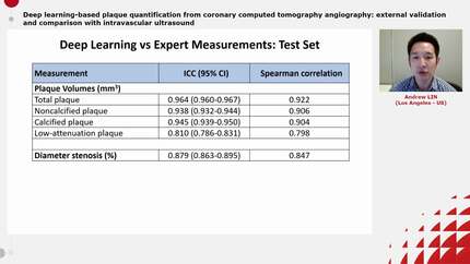
Optical coherence tomography (OCT) provides excellent resolution at the cost of shallow penetration, limiting its ability for plaque burden (PB) assessment. A deep learning (DL) algorithm was therefore developed to provide fully automated assessment of PB and vessel size from OCT. We aimed to perform a head-to-head comparison of AI-powered OCT vs. intravascular ultrasound (IVUS) for PB quantification, using histopathology as the reference standard.
Ten post-mortem human hearts were harvested. All 29 major epicardial coronary arteries were perfused and underwent OCT and IVUS pullbacks. After imaging, the arteries were serially sectioned at 1-mm intervals for histopathology. Lumen and vessel contours were derived fully automatically from OCT using the DL algorithm and semi-automatically from IVUS. Histopathological measurements were obtained by experienced analysts. Statistical analyses accounted for within-patient clustering. A total of 852 paired OCT-IVUS histopathology cross-sections were obtained. AI-powered OCT showed strong correlations with histopathology for the assessment of lumen area (
In this histopathology validation study of 10 post-mortem human hearts, AI-powered OCT enabled fully automated plaque burden quantification and correlated strongly with histopathology, with no statistically significant difference between the OCT–histopathology and IVUS–histopathology correlations.
Contributors

Ruobing Dai
Author
Xu Yu
Author
Miao Chu
Author
Su Zhang
Author

Sean Hynes
Author
Yining Wang
Author
William Wijns
Author
Shengxian Tu
Author
You may be interested in