Dynamic left main occlusion after aortic valve surgery: recognizing extrinsic compression by aortic root pseudoaneurysm—a case report
European Heart Journal - Case Reports


Abstract
Extrinsic compression of the left main (LM) coronary artery by an aortic root pseudoaneurysm is an exceedingly rare but life-threatening cause of acute coronary syndrome following aortic valve surgery. Multimodality imaging is essential for diagnosis and surgical planning.
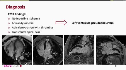
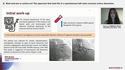
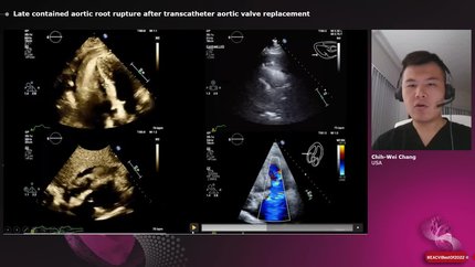
A 52-year-old male underwent mechanical aortic valve replacement and aortic root reconstruction for infective endocarditis complicated by a 23 mm aortic root abscess. Following initial recovery, he re-presented 2 months later with persistent chest pain on a background of 10 days of intermittent symptoms, with anterior-lateral ischaemic changes on electrocardiography (ECG). Computed tomography (CT) aortography excluded dissection but was not formally reported. In the catheterization laboratory, fluoroscopy revealed abnormal prosthetic valve rocking consistent with dehiscence. Coronary angiography demonstrated absent antegrade flow in the left anterior descending artery, impaired flow in the left circumflex artery, and a pulsatile, systole-predominant LM lesion strongly suggesting extrinsic compression. No percutaneous intervention was performed. Urgent formal CT review confirmed a 5.2 × 4.4 × 4.0 cm left ventricular outflow tract pseudoaneurysm with the LM coursing within its fold and apparent systolic occlusion. Serial ECGs showed gradual improvement, supporting a dynamic mechanism. After multidisciplinary review, the patient underwent successful composite graft replacement and pseudoaneurysm repair at a quaternary centre, with full recovery at 4-month follow-up.
This case illustrates three key lessons: the dynamic, systole-predominant angiographic hallmark of extrinsic LM compression distinguishes it from fixed atherosclerotic disease and prevents inappropriate percutaneous intervention; dynamic angiography and CT play complementary roles, and single-phase systolic CT may, in this setting, overestimate the severity of LM occlusion; and early referral to a specialist quaternary surgical centre is essential for definitive repair.
Contributors

Sherif Seif
Author
Royal Preston Hospital Preston , United Kingdom of Great Britain & Northern Ireland
Babatunde Kukoyi
Author
Abdalla Mujahid
Author
Omar Nawaytou
Author
Karim Ratib
Author
Krishnaraj Rathod
Author
Frederic De Roeck
Author
Giovanni Martino
Author
Deepti Ranganathan
Author
You may be interested in