Non-invasive left ventricular pressure-volume loops from cardiovascular magnetic resonance imaging and brachial blood pressure: validation using pressure catheter measurements
European Heart Journal - Imaging Methods and Practice


Abstract
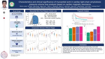
Left ventricular (LV) pressure-volume (PV) loops provide gold-standard physiological information but require invasive measurements of ventricular intracavity pressure, limiting clinical and research applications. A non-invasive method for the computation of PV loops from magnetic resonance imaging and brachial cuff blood pressure has recently been proposed. Here we evaluated the fidelity of the non-invasive PV algorithm against invasive LV pressures in humans.
Four heart failure patients with EF < 35% and LV dyssynchrony underwent cardiovascular magnetic resonance (CMR) imaging and subsequent LV catheterization with sequential administration of two different intravenous metabolic substrate infusions (insulin/dextrose and lipid emulsion), producing eight datasets at different haemodynamic states. Pressure-volume loops were computed from CMR volumes combined with (i) a time-varying elastance function scaled to brachial blood pressure and temporally stretched to match volume data, or (ii) invasive pressures averaged from 19 to 30 sampled beats. Method comparison was conducted using linear regression and Bland-Altman analysis. Non-invasively derived PV loop parameters demonstrated high correlation and low bias when compared to invasive data for stroke work (R2 = 0.96,
Pressure-volume loops can be precisely and accurately computed from CMR imaging and brachial cuff blood pressure in humans.
Contributors

Per M Arvidsson
Author
Peregrine G Green
Author
William D Watson
Author

Mayooran Shanmuganathan
Author
Wycombe Hospital High Wycombe , United Kingdom of Great Britain & Northern Ireland
Einar Heiberg
Author
Giovanni Luigi De Maria
Author
Håkan Arheden
Author
Neil Herring
Author
University of Oxford Oxford , United Kingdom of Great Britain & Northern Ireland
You may be interested in