Characterization of intraplaque haemorrhage in statin-treated patients with stable coronary artery disease: multi-modality imaging with non-contrast T1-weighted cardiac magnetic resonance, optical coherence tomography, and near-infrared spectroscopy

European Heart Journal - Imaging Methods and Practice

7 August 2026
Organised by: Logo
ESC Journals CORONARY ARTERY DISEASE, ACUTE CORONARY SYNDROMES, ACUTE CARDIAC CARE IMAGING Cardiac Magnetic Resonance (CMR) Hybrid and Fusion Imaging PREVENTIVE CARDIOLOGY Risk Factors and Prevention

Abstract

AbstractAims

Plaque-to-myocardium ratio (PMR) ≥1.0 on cardiac magnetic resonance (CMR) has been histologically validated to correspond to intraplaque haemorrhage. Despite the use of statins, PMR values remained above 1.0. Detailed plaque features exhibiting residual intraplaque haemorrhage despite statin therapy remains unknown.

Objectives

To characterize features of plaques exhibiting PMR ≥1.0 in statin-treated patients with CAD.

Methods and results

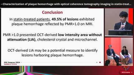
Target (n = 161) and non-target (n = 142) lesions in 121 statin-treated stable CAD patients were imaged by CMR, near-infrared spectroscopy (NIRS)/intravascular ultrasound (IVUS), and optical coherence tomography (OCT) (NCT04864171). NIRS/IVUS-derived measures, OCT-derived low-intensity area without attenuation (LIA), and the arc of LIA were compared between lesions with and without PMR ≥1.0. PMR ≥1.0 was observed at 34.2% and 19.7% of target and non-target lesions, respectively. Target lesions with PMR ≥1.0 presented greater percent atheroma volume (PAV) and higher maxLCBI4mm with greater frequencies of layered plaques, cholesterol crystals, and LIA (P < 0.001 for all). Multivariable analysis demonstrated PAV [odds ratio (OR) = 1.05, 95% confidence interval (CI) = 1.01–1.09, P = 0.006], layered plaque (OR = 3.17, 95% CI = 1.03–9.78, P = 0.045), cholesterol crystal (OR = 3.79, 95% CI = 1.30–11.03, P = 0.015), and LIA arc (OR = 1.04, 95% CI = 1.01–1.06, P < 0.001) but not maxLCBI4mm (OR = 1.001, 95% CI = 0.999–1.003, P = 0.227) as predictors of PMR ≥1.0 at target lesions. Notably, the frequency of PMR ≥1.0 increased to 92.0% in target lesions exhibiting all plaque features (PAV ≥ 62.1%, layered plaque, cholesterol crystal, LIA arc ≥18.0°). In non-target lesions, PAV ≥ 53.1%, cholesterol crystal and LIA arc ≥16.5° predicted PMR ≥1.0. A clustering of these features elevated the frequency of PMR ≥1.0 (P < 0.001).

Conclusion

Statin-treated patients still harboured PMR ≥1.0. PAV, LIA, and cholesterol crystal predicted PMR ≥1.0 at both target and non-target lesions. Clustering of these plaque characteristics may enable to better identify intraplaque haemorrhage in vivo.

Contributors

Yu Kataoka
Yu Kataoka

Author

National Cerebral & Cardiovascular Center Suita , Japan

Kentaro Mitsui
Kentaro Mitsui

Author

National Cerebral & Cardiovascular Center Suita , Japan

Kota Murai
Kota Murai

Author

National Cerebral and Cardiovascular Center Hospital Osaka , Japan

Yasuhide Asaumi
Yasuhide Asaumi

Author

National Cerebral & Cardiovascular Center Suita , Japan