One-click co-registered T2 mapping and dual black- and bright-blood late gadolinium enhancement MRI for comprehensive assessment of myocardial injury after acute STEMI
European Heart Journal - Imaging Methods and Practice


Abstract
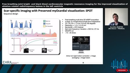
In acute ST-segment elevation myocardial infarction, ischaemia and reperfusion lead to a cascade of myocardial injury that can be characterized by cardiac magnetic resonance (CMR) imaging, including coagulation necrosis, oedema, papillary muscle damage, microvascular obstruction, and intramyocardial haemorrhage. Conventional CMR protocols require multiple sequences to be performed and complicated analysis. This study evaluates SPOT-MAPPING, a sequence that acquires co-registered T2 maps and dual bright- and black-blood late gadolinium enhancement (LGE) images in a single scan.
SPOT-MAPPING employs a single-shot, ECG-triggered 2D sequence alternating between bright- and black-blood LGE imaging with varying T2 weightings. We prospectively enrolled 20 STEMI patients undergoing CMR at 1.5 T within 4–7 days post-emergent coronary intervention. SPOT-MAPPING’s segmentation times and reproducibility of myocardial injury markers (oedema, scar size, transmurality, papillary muscle damage) were assessed against conventional T2 mapping and phase-sensitive inversion recovery (PSIR). SPOT-MAPPING halved left ventricular wall segmentation time (∼3 min) while maintaining high reproducibility for oedema, scar size, and transmurality (ICC > 0.8). It improved papillary muscle damage detection over PSIR (8 vs. 3 patients) and showed comparable T2 values with conventional T2 mapping (remote: 45.0 ± 3.6 ms vs. 45.9 ± 3.7 ms,
SPOT-MAPPING offers a time-efficient and reproducible CMR method for myocardial injury assessment post-STEMI.
Contributors


Victor de Villedon de Naide
Author
Thaïs Génisson
Author
Kalvin Narceau
Author
Théo Richard
Author
Konstantinos Vlachos
Author
Guido Caluori
Author
Claire Bazin
Author
Soumaya Sridi
Author
Ilyes Benlala
Author
Gael Dournes
Author
Maxime Sermesant
Author
Michel Montaudon
Author
Pierre Jaïs
Author
Matthias Stuber
Author
Hubert Cochet
Author
You may be interested in