Sex-specific shape models to disentangle sex effects and age-associated anatomical variation in biventricular cardiac anatomy
European Heart Journal - Imaging Methods and Practice


Abstract
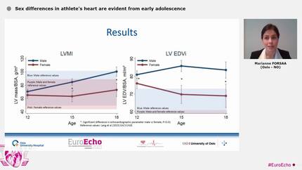
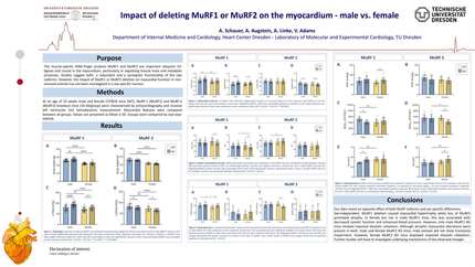
Age- and sex-related differences in cardiac anatomy are well established using conventional cardiovascular magnetic resonance biomarkers, but their representation in 3D ventricular shape remains unclear. In pooled statistical shape models, leading modes may be dominated by between-sex anatomical variation, potentially obscuring within-sex age-associated variation. This study evaluated whether sex-specific statistical shape models provide a more interpretable representation of age-associated ventricular variation than a sex-agnostic model.
Biventricular meshes were reconstructed from cine cardiovascular magnetic resonance (CMR) images in a cross-sectional screened asymptomatic Swiss adult cohort aged 18-85 years. End-diastolic anatomies from 185 male and 187 female participants were used to construct statistical shape models for the pooled cohort and separately by sex. Shape modes were interpreted using anatomical visualization, variance decomposition, associations with conventional demographic, anthropometric, and CMR biomarkers.
The first pooled model mode explained 31% of total shape variance, compared with 22% and 19% for the first male- and female-specific modes. In the pooled model, 57% of first-mode variance was attributable to between-sex differences. Age-associated variation in the pooled model was distributed across the first two modes, whereas sex-specific models organized age-associated variation within each sex. In males, the strongest adjusted age association occurred in the second sex-specific mode, which correlated with ventricular volume and mass. In females, age-associated variation was concentrated in the first mode and was inversely associated with ventricular volumes and myocardial mass.
These findings support sex-specific anatomical reference models for capturing within-sex cardiac ageing structure.
Contributors

Stefano Buoso
Author
Valery L Visser
Author
Pia Leuders
Author
Tobias Kudelka
Author
Maximilian Fuetterer
Author
Christina Spengler
Author

Sebastian Kozerke
Author
You may be interested in