Clinical validation of in-silico pace-mapping for non-invasive guidance in ablation planning
EP Europace Journal


Abstract
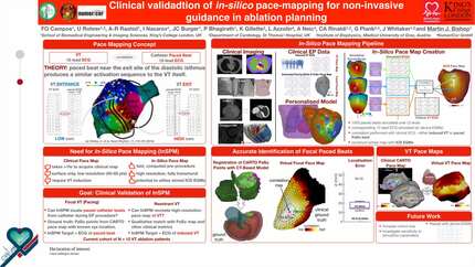
In-silico pace-mapping uses a patient-specific computational model alongside the patient’s ECG to generate a 'virtual' fully 3D pace map for guiding ablation therapy. Whilst a promising pre-procedural tool to optimize treatment, its clinical effectiveness remains to be validated through further clinical data.
To assess the ability of in-silico pace-mapping in identifying the site of focal origin during clinical pacing.
High-resolution contrast computer tomography (CT) and late enhancement cardiac wideband MRI of three patients were obtained pre-ablation procedure and used to construct personalized torso-ventricular computational models (Fig. A). Infarct anatomy (scar and border zone) was obtained either from commercially available segmentation software (ADAS3D) (n=2) or voltage data and mapped onto the torso-ventricular models (n=1) (Fig. B). ECGs from body-surface locations (Fig. C) were obtained during pace mapping procedure, and their respective pacing sites (N~16 per patient) exported from the mapping system (CARTO, Biosense Webster) and used as ground truth. In-silico pace-mapping involved pacing the heart at 1000 random sites on the surface of the left ventricle and computing respective ECGs. Location of ECG electrodes on the patient’s torso was inferred through registered fluoroscopy images to ensure close match. Correlation analysis compared clinically-acquired and simulated ECGs for each of the 1000 pacing sites, constructing virtual pace maps (Fig. D). The average of the top 10 coefficients across leads (ccm10) was calculated. The distance (d) between the pacing electrode and the location with the strongest correlation was determined (Fig. D). The median and interquartile range of d across all patients and pacing locations (N=51) was calculated.
Fig. D presents the pace map obtained for one patient paced a representative location (blue star). The strongest correlation point (ccm10=0.716) is marked by a red star with a distance to the ground-truth (blue star) of 7.5mm. The median distance for this patient across all N=12 pacing sites was d = 13.7mm (IQR: 11.0-27.6) compared to d = 24mm (IQR: 16.1-28.4, N=18) and d = 43mm (IQR: 24.2-53.5, N=21) for the other two patients. The spatial precision across all patients (N=51), shown in Fig. E, was 26.6mm (IQR: 16.4-16.4).
This work demonstrated the value of in-silico pace mapping in determining the origin of ventricular pacing in patient-specific ventricular-torso models, offering a rapid, risk-free pre-procedure testbed. However, further validation on a larger patient cohort is necessary to enhance robustness and broaden clinical applicability.
Contributors

F O Campos
Author
U Rohrer
Author
A R Rashid
Author
L Nazarov
Author
J C Burger
Author
P Bhagirath
Author
L Azzolin
Author
A Neic
Author
C A Rinaldi
Author
G Plank
Author
J Whitaker
Author
M J Bishop
Author
You may be interested in