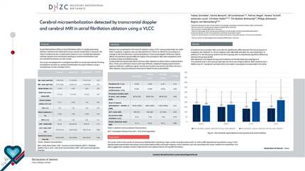
Comparison of cerebral micro-embolization detected by transcranial doppler examination between radiofrequency ablation and pulsed field ablation for atrial fibrillation
EP Europace Journal


Abstract
Cerebral ischemia is a rare but severe complication after catheter ablation (CA) of atrial fibrillation. Pulsed Field Ablation (PFA) is a promising alternative nonthermal ablation method establishing pulmonary vein isolation via irreversible electroporation and is believed to reduce overall complications. The incidence of ischemic lesions on cerebral MRI after PFA has been described as low as 3%, but as PFA systems differ considerably and more safety data is needed. Real-time assessment of microembolic signals (MES) measured by transcranial doppler (TCD) may help to identify critical procedural steps associated with periprocedural stroke. Therefore, we sought to compare the incidence of MES assessed by TCD during different procedural steps between PFA and radiofrequency ablation (RFA).
Consecutive patients who met the following inclusion criteria were prospectively recruited at the DHZC, Campus Benjamin Franklin, between 2022 and 2024: (a) first-time CA of paroxysmal or persistent atrial fibrillation (b) no anticipated additional left atrial ablation.
TCD was performed using a pulsed-wave 2-MHz Doppler probe fixed to the patient’s head with the DiaMon (DWL) system. To exclude artifacts, each detected MES was manually controlled by a neurologist after the procedure was completed.
MES were assigned to the following procedural steps: (1) transseptal puncture, (2) introduction of catheters (3) electroanatomical mapping, (4) ablation and (5) removal of the catheters.
All patients gave written informed consent and underwent continuous TCD examination during CA. Additionally, postprocedural NIHSS (National Institutes of Health Stroke Scale) scores were taken.
In total, 32 patients were analyzed and 26 patients were included for statistical analysis (see figure 1). CA was successfully performed with single transseptal puncture in all patients. MES measuring was obtained using a unilateral window. PFA had significantly higher MES-counts, especially during ablation, compared to RFA (see table 1).
Two complications occurred in the PFA group (one postinterventional ischemic stroke and one transient phrenic nerve palsy) while no complication was seen in the RFA group (p=0.065). In the patient with ischemic stroke, the MES-count was not elevated above average (n=319 in total, see table 1).
MES-count varies significantly between RFA and PFA. To our knowledge, the observed MES for both ablation technologies rank among the lowest reported in literature. Further studies including pre- and postprocedural MRI are needed to confirm the findings.
Contributors

T Schreiber
Author
P Attanasio
Author
N Martins
Author
U Landmesser
Author
P Nagel
Author
V Tscholl
Author
M Huemer
Author
J Lucas
Author
C Nolte
Author
T Braemswig
Author
R Von Rennenberg
Author
You may be interested in